
Types of Imaging:
- Confocal
- Live Cell
- Widefield
- Multiphoton
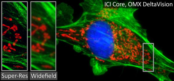
- Super Resolution
Booking:
Bookings can be made online for the scopes through PPMS. To view the calendars, please click here. Pediatric users are also welcome to use microscopes at any of the ICI locations.
A small sampling of our available microscopes in the HSRB I/II complex is below. For a more exhaustive list, FAQs, pricing/rates, subsidy information, and further, please visit our Emory Integrated Core Facility page.
HSRB I & II Microscopes
Nikon CSU-W1 SoRa Spinning Disk / HSRB I
The CSU-W1 SoRa is a confocal field scanning spinning disk system and a confocal super resolution system in a single package. We offer dry as well as water immersion lenses for high resolution imaging with fixed and live cell.
- Fast and gentle spinning disk confocal
- two disks, one for confocal, one for super resolution
- easy switch between the two disks
- Four laser lines [nm]: 385, 475, 550, and 621
- Z-stacks, timelapse, multi-point imaging, tiling
- Live-cell imaging with stage top incubator
Leica STELLARIS 8 / HSRB II
- White light laser, extended into the near IR allowing for the application and separation an extended range of spectrally overlapping fluorophores.
- Point scanning inverted confocal
- Galvano and resonant scanners
- 5 detectors: 3 Power HyD-S and 2 Power HyD-X
- Tokai Hit stage top incubator for CO2 and temperature control
- Whitlight lasers, 440-790
- Navigator software for tiling
- TauSense for FLIM
- LIGHTNING for optimal extraction of image details and maximum resolution
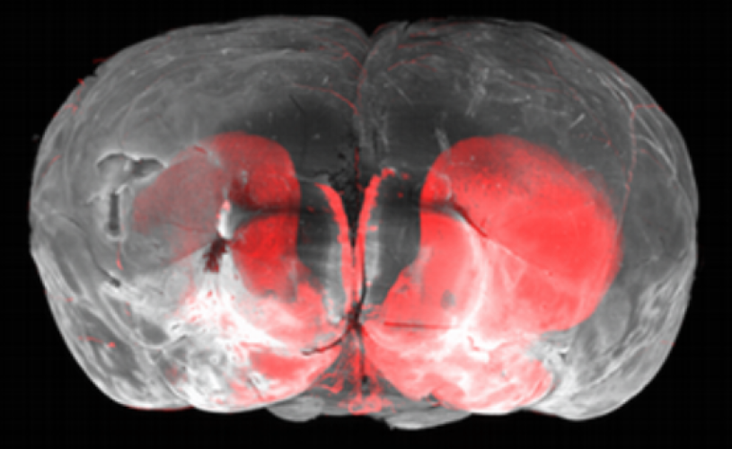
Miltenyi UltraMicroscope Blaze Lightsheet / HSRB II
A fully automated light sheet microscope for imaging large or multiple cleared samples at subcellular resolution.
- Super resolution achieved through the clearing of tissue, organs or small animals allowing for complete 3D mapping at single cell resolution.
Leica Thunder Widefield / HSRB II
3D live cell wide field microscope. The Thunder imager helps to maintain optimal physiological conditions by minimizing photobleaching, providing high performance imaging.
- fully motorized DMi8 microscope
- highly sensitive K8 camera
- multi-line, high-intensity fluorescence LED light source
- Live cell imaging with a Tokai Hit stage top incubator
- Computational clearing to minimize out of focus blur
Meet the Team

April Reedy, PhD
Technical Director
