
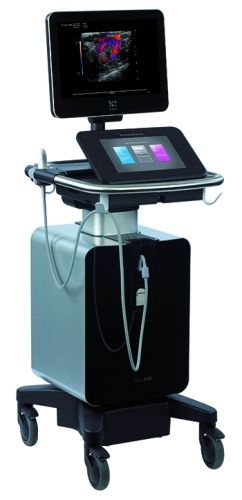
Ultrasound and Your Research Toolkit
Neurology
Our first webinar in our multi-part series gives the basics of ultrasound and delves into specifics on applications in neurology. AP's Technical Director Rebecca is joined by Isabel Newsome, PhD of Fujifilm VisualSonics.
High-Frequency Ultrasound Imaging
Anatomical Motion mode allows the user to steer the sample volume to any angle rather than containing it strictly in a vertical position.
2D and 3D Color Flow acquisition for blood flow velocity analysis using advanced digital signal processing techniques.
- Perfusion Analysis using destruction/reperfusion quantification.
- Biomarker quantification tools when using Vevo MicroMarker Target-Ready Contrast Agents.
Acquisition can be triggered at one or two points in the cardiac cycle.
- Improved and repeatable LV function analysis.
- LV trace can be generated automatically with one button (Auto LV).
Single line acquisition that allows high temporal resolution for LV functional analysis.
2D and 3D Power Doppler acquisition for the visualization of slow velocity flow and relative quantification of smaller vessels using advanced digital sign processing techniques.
- Quantification Software for perfusion and targeted signal analysis.
- Perfusion parameters (including amplitude and time) derived from a curve-fitting algorithm for bolus kinetics and replenishment kinetics following destruction reperfusion.
- Spatial rendering is available in the form of parametric maps.
- Data can be exported in .tiff, .bmp, .wmv files and. tsv formats.
- Analysis suite provides tools for myocardial strain, strain rate, and time-to-peak analysis.
- The feature also includes a dynamic assessment of myocardial velocity and displacement.
- The analysis suite provides tools for vascular strain, strain rate, and displacement.
- This feature also includes Pulse Propagation Velocity assessment.
Click here to see a list of custom-designed linear array transducers for Vevo 3100.
Meet the Team

David Archer, PhD
Scientific Director, Pediatric+Winship Flow Cytometry Core and Animal Physiology Core